History
The history is arguably the most important aspect of the whole examination. It is from the history that the nature of the problem is determined and possible treatments are decided upon (Refshauge and Gass, 1995). Patients with recurrent anterior shoulder dislocations commonly complain of the signs and symptoms listed on the previous page.
Physical Assessment
A physical assessment should be performed by a trained medical professional and may comprise of any of the following:
- Observation - Muscle bulk of the shoulder girdle, anatomical abnormalities, posture, swelling or bruising
- Palpation - Feeling for tenderness around the shoulder and surrounding structures or abnormalities compared to the unaffected side
- Range of motion (ROM) - Investigation of the movement within the joint
- Functional movements - Looking for quality of shoulder movement and identifying any factors
- Neurological (nerve) / vascular (blood flow) testing - The clinician will test to see if your shoulder has normal strength, sensation and co-ordination
-
Special tests - may be used to test specific structures or to determine whether or not a particular type of condition or diseases is present (Magee, 2008)
The clinician may perform a number of tests to examine :
Commonly used special tests to assess anterior (forward) instability associated with a recurrent anterior shoulder dislocation are:
| 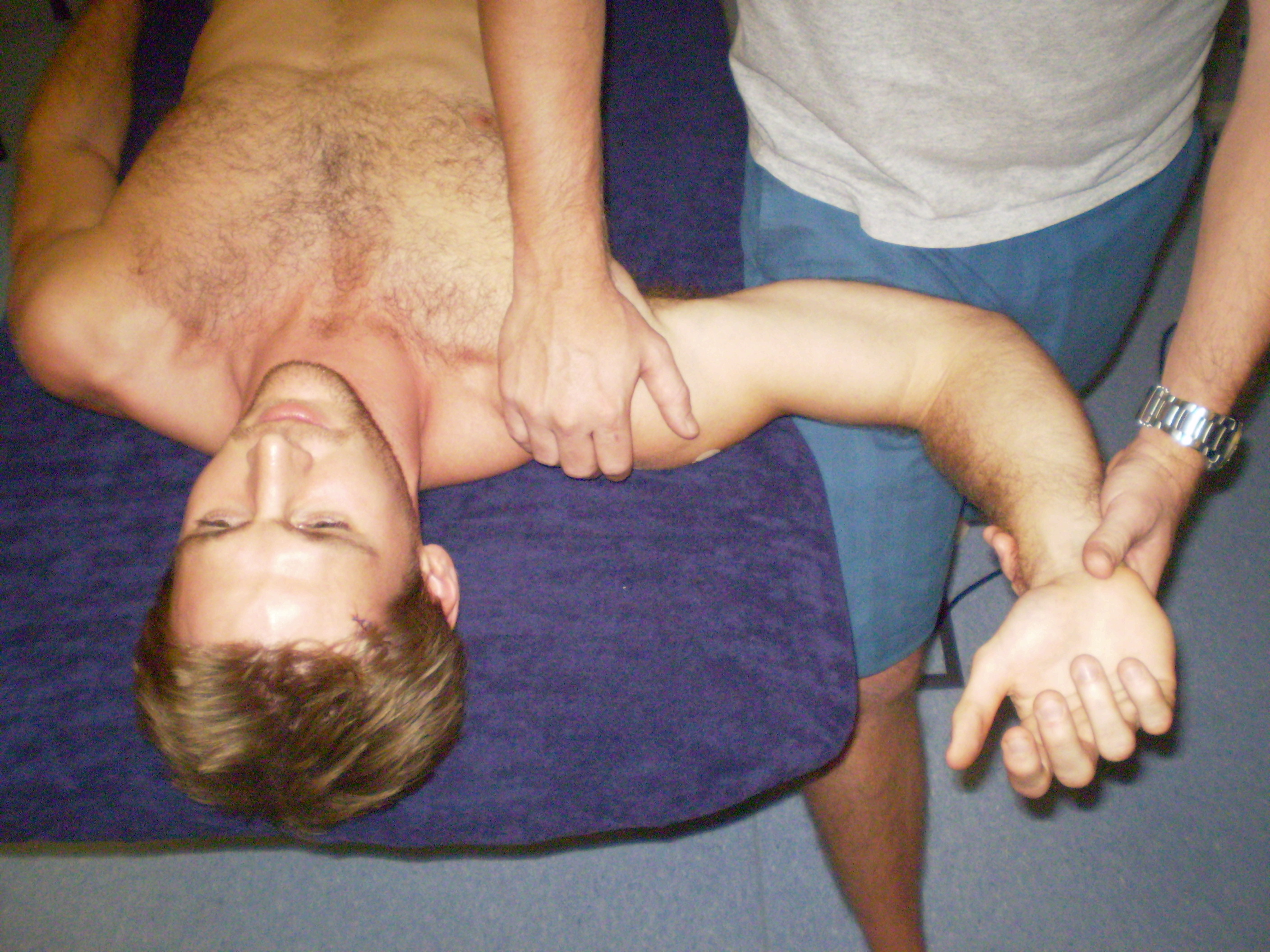 Apprehension relocation: Patient is placed into lying position. Arm is supported by the clinician and turned outwards. Some discomfort may be experienced. If so, a slight pressure to the top of the arm bone will be applied. If apprehension is decreased, the test is postive for anterior (forward) instability. |
To read more about specific shoulder tests a helpful link is shoulderdoc.co.uk
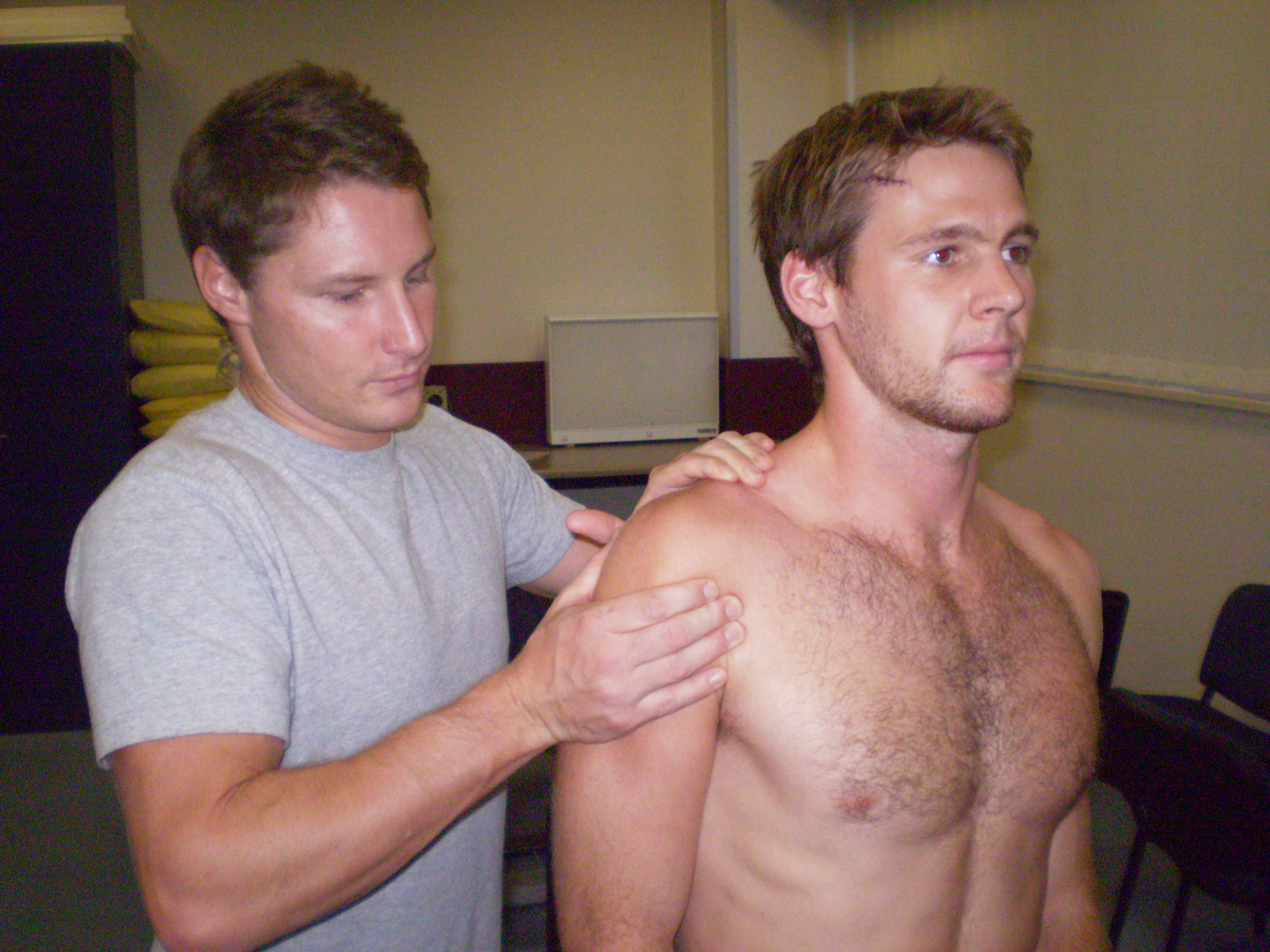 | 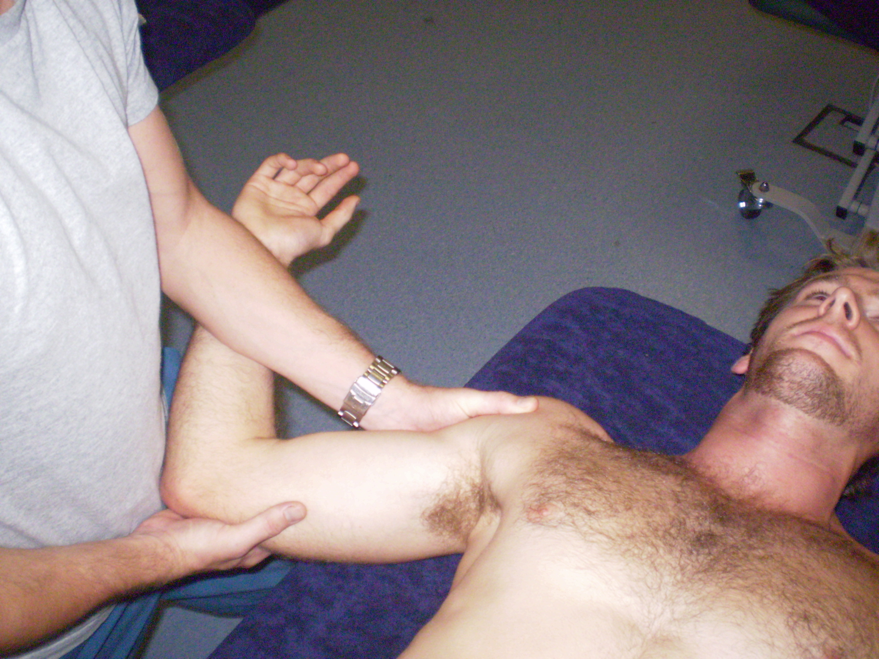 |
Load and shift: The arm is lifted into its neutral position. The patient will feel the top of the arm bone moving forwards and backwards to provide the therapist with the degree of movement present within the joint. Greater movement or onset of symptoms in the injured arm may indicate a positve test. | Anterior draw: the clinician will support the arm in a turned out position. The patient will feel the arm being moved towards the ceiling in a number of different positions. In a positive test symptoms may be reproduced or excessive movement may occur. |
Other investigations Medical imaging may be used to identify tissue damage as well as rule out other possible pathologies. Some associated pathologies are mentioned on the signs and symptoms page. X-ray: Multiple X-rays must be taken prior to reduction to determine the direction and extent of the dislocation. It may also detect the existence of associated fractures and possible barriers to relocation. MRI - Gives information about structures that can not be seen as clearly with an X-ray such as damage to ligaments, tendons, cartilge or labrum which commonly occur in repeated (recurrent) shoulder dislocations. MRI may also be used to diagnose unexplained shoulder pain. | 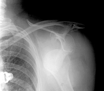 X-ray of anteriorly dislocated left shoulder (looking from the front) |